
Muscle Contraction
The process of muscular contraction occurs over a number of key steps, including:
-
Depolarisation and calcium ion release
-
Actin and myosin cross-bridge formation
-
Sliding mechanism of actin and myosin filaments
-
Sarcomere shortening (muscle contraction)
1. Calcium Ion Release
-
Contraction of a muscle fibre begins when an action potential from a motor neuron triggers the release of acetylcholine into the neuromuscular junction (motor end plate)
-
Acetylcholine initiates depolarisation within the sarcolemma, which is spread through the muscle fibre via membrane invaginations called T tubules
-
Depolarisation causes the sarcoplasmic reticulum to release stores of calcium ions, which play a pivotal role in initiating muscular contractions


2. Cross-Bridge Formation
-
Muscle fibres contain long myofibrils composed of a protein complex of actin and myosin (myofilaments)
-
On actin, the binding sites for the myosin heads are covered by a blocking complex (troponin and tropomyosin)
-
Calcium ions bind to troponin and reconfigure the complex, exposing the binding sites that were being blocked by tropomyosin
-
With the binding sites now exposed, the myosin heads form a cross-bridge with the actin filaments
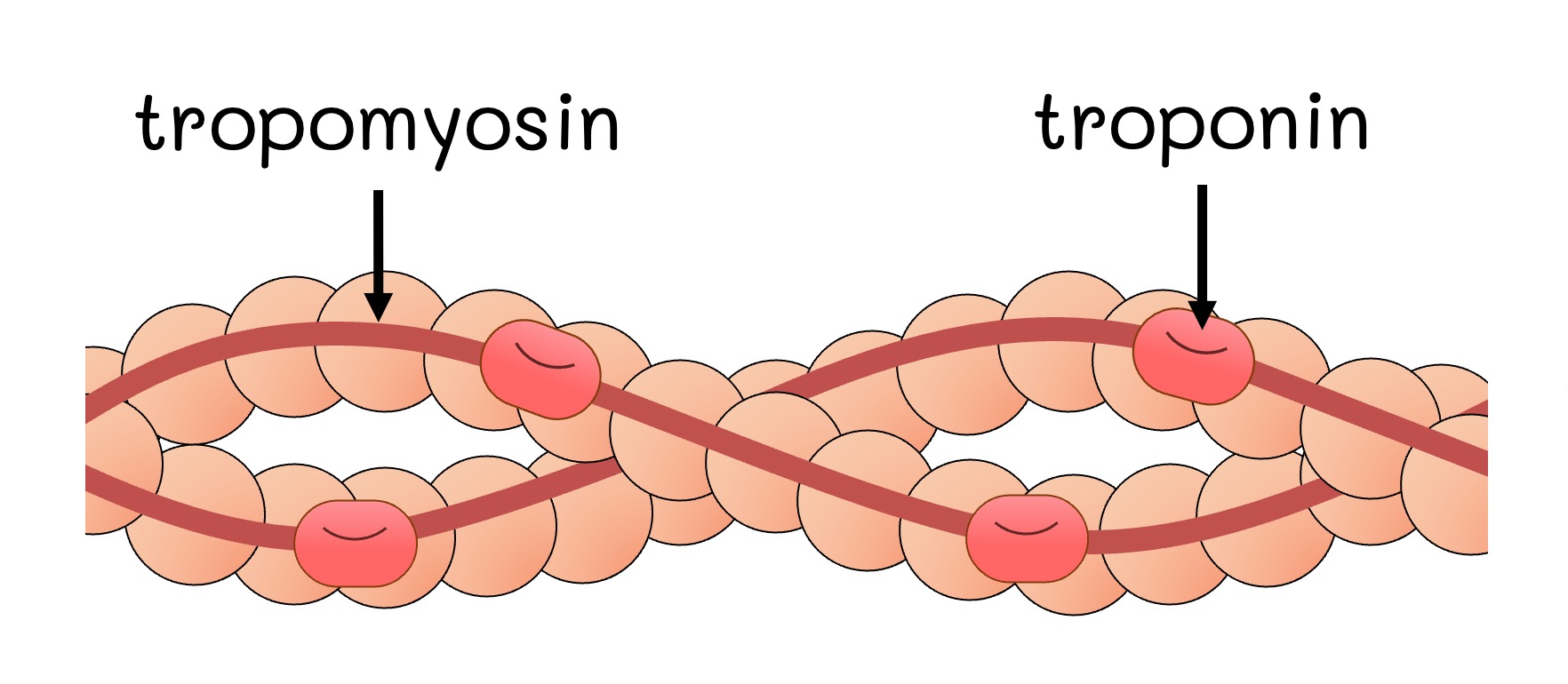
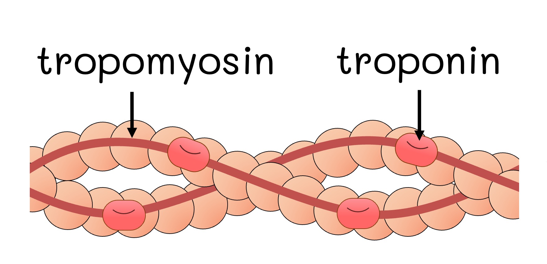
Covered
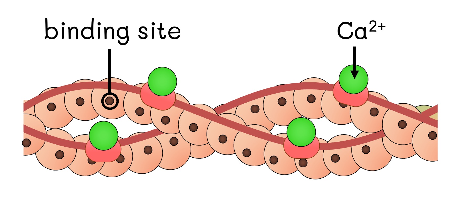
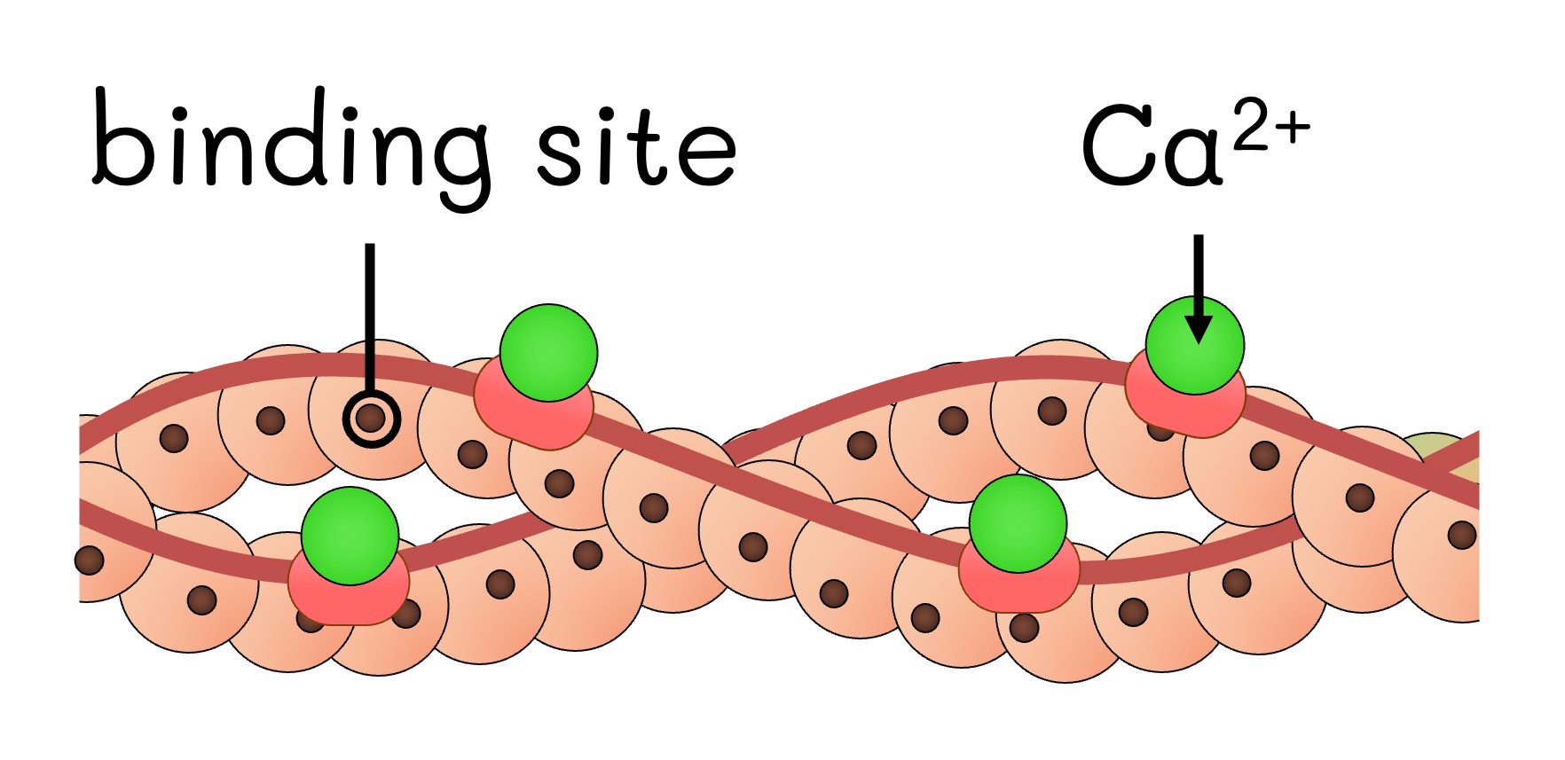
Exposed
3. Sliding Mechanism
-
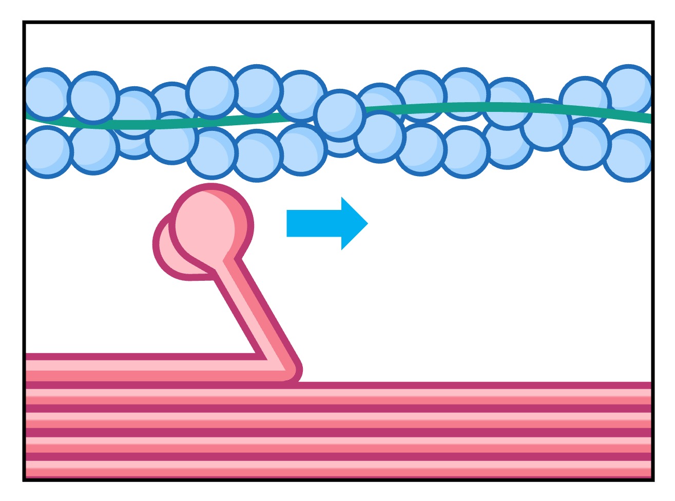
ATP binds to the myosin head, breaking the cross-bridge between actin and myosin
-
ATP hydrolysis causes the myosin heads to change position and swivel, moving them towards the next actin binding site
-
The myosin heads bind to the new actin sites and return to their original conformation
-
This reorientation drags the actin along the myosin in a sliding mechanism
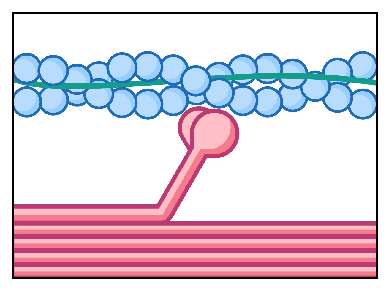
Cross-bridge
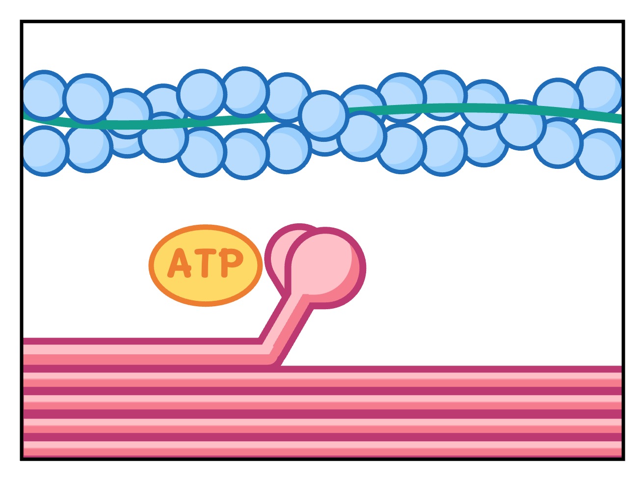
ATP binding
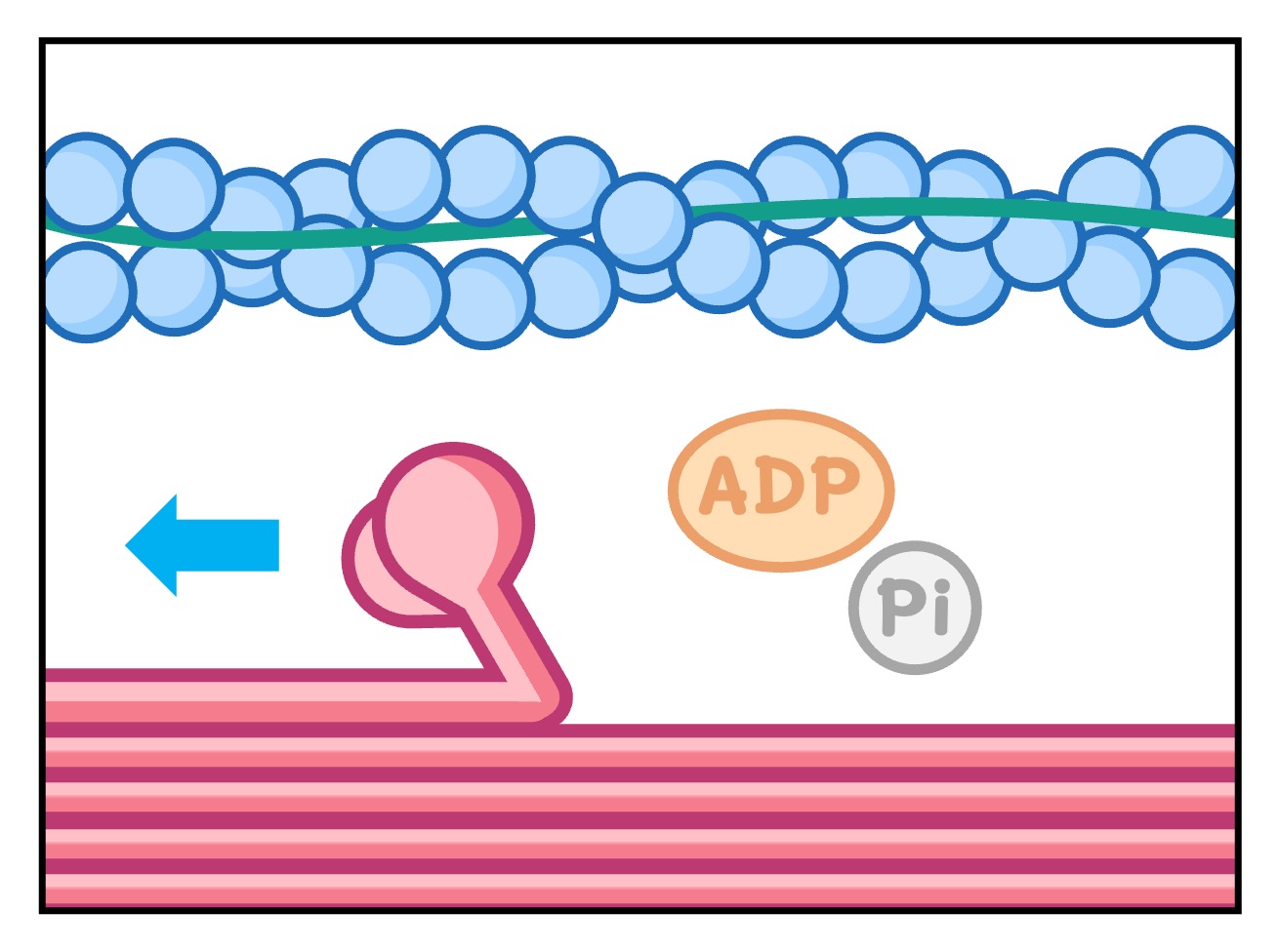
Reconfiguration
Sliding Filament
4. Sarcomere Shortening
-
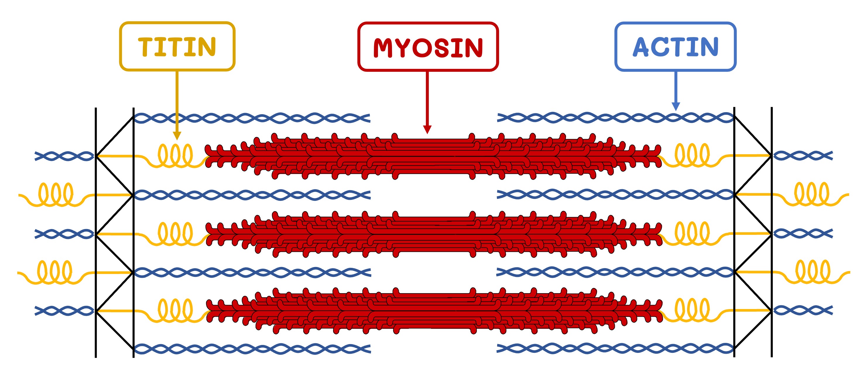
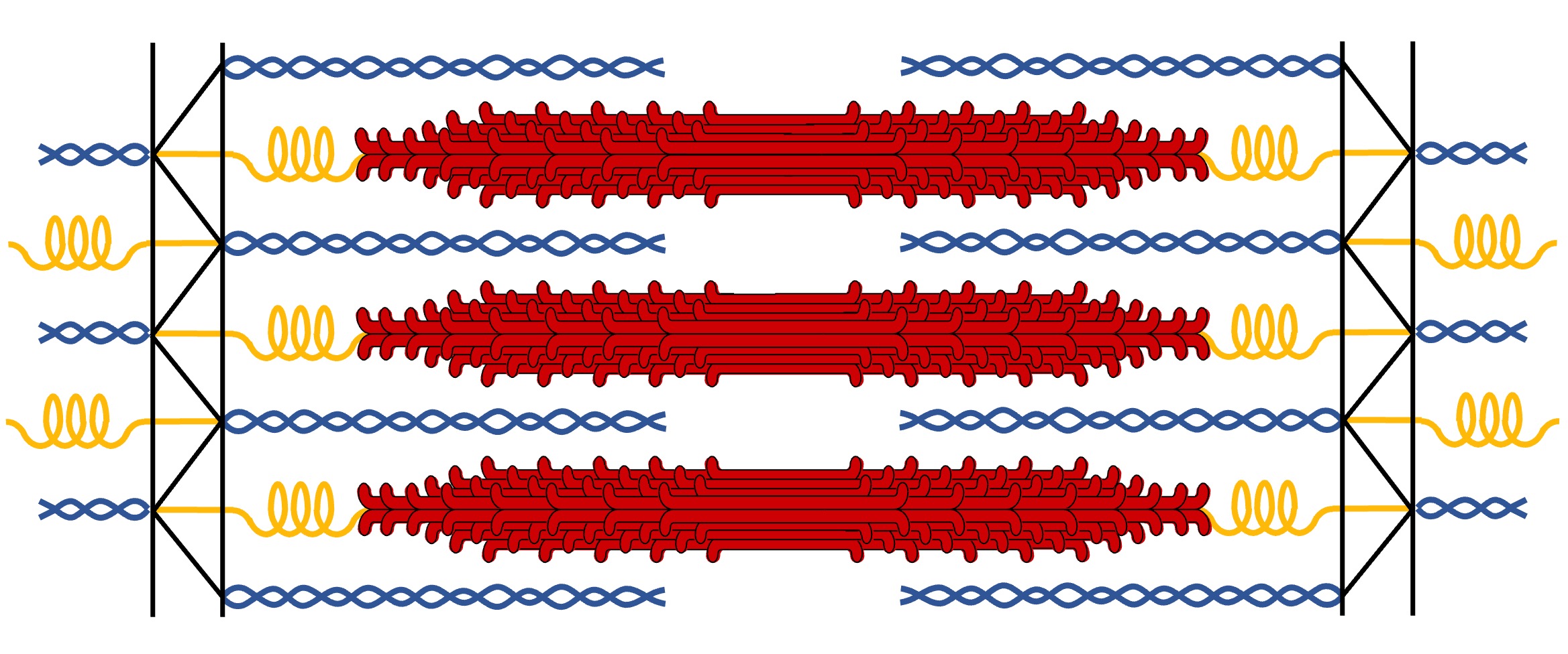
The myofilaments form structural units called sarcomeres (each sarcomere functions as a contractile unit)
-
Actin filaments are attached directly to Z lines, while myosin filaments are anchored to Z lines via the protein titin
-
As actin filaments slide along the myosin, the actin pulls the Z lines closer together, shortening the sarcomere
-
As the individual sarcomeres become shorter in length, the muscle fibres as a whole contracts
Muscle Relaxation
Muscle relaxation relies on a protein called titin, which connects the myosin filaments to the Z line
-
The many folds of the titin molecule gives it spring-like properties, allowing it to store elastic potential energy
-
Titin can either be compressed (during muscle contraction) or stretched (during muscle relaxation)
-
The titin can then recoil and convert its elastic potential energy into kinetic energy, moving Z lines apart (if compressed) or closer together (if stretched)
The stretching of a muscle requires a second muscle, as muscles can only work by contracting
-
Hence, skeletal muscles work in antagonistic pairs – when one muscle contracts, the other muscle relaxes
-
When titin is compressed in one muscle, it will lengthen in the antagonistic muscle (gaining elastic potential energy in both)


